


 返回顶部
返回顶部

光解仪 超低折扣,只需5折!
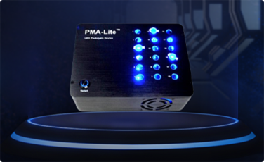
PMA-Lite™ LED Photolysis Device
PMA-Lite LED 光解装置是一种轻型 LED 灯箱,专为在活力 PCR 应用中对PMAxx或PMA处理过的样品进行最佳光解而设计。
查看产品
PMA 实时 PCR 活力试剂盒
设计用于对肠道沙门氏菌、单核细胞增生李斯特氏菌、金黄色葡萄球菌、MRSA、大肠杆菌、嗜肺军团菌、结核分枝杆菌、大肠杆菌O157:H7进行活力 PCR 的试剂盒
PMA (Propidium Monoazide)
一种光反应 DNA 结合染料,通常用于 细菌、病毒和真菌等微生物的活力 PCR (v-PCR)。
PMAxx™ Dye, 20 mM in H2O
活力 PCR 染料是流行的活力染料单叠 氮化丙啶 (PMA) 的新改进版本。
产品分类

淀粉样蛋白和神经变性染色
抗体和蛋白质标记试剂盒

细胞凋亡和活力测定

缓冲液、附件和背景抑制剂

钙指示剂和螯合剂

癌症研究

细胞和细胞器染色

细胞周期分析

显色底物

DNA 凝胶染色剂和配件

酶底物

设备

流式细胞术

荧光显微镜

荧光蛋白、核苷酸和其他偶联物

HRP 和碱性磷酸酶偶联物

体内成像

萤光素酶报告基因检测

镁、氯化物、pH、锌和其他指标

微生物学

神经科学

核复染剂

核酸定量和提取

其他 qPCR 试剂

PCR 和 DNA 扩增

一抗

活性 CF® 染料、其他活性染料和生物素化试剂

一氧化氮 (NO) 和活性氧 (ROS) 试剂

二抗、抗标签抗体和链霉亲和素偶联物

超分辨率成像

Western和蛋白质分析

qPCR 染料

qPCR 预混液

免疫学抗体
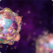
技术亮点:ExoBrite EV 膜染色试剂盒
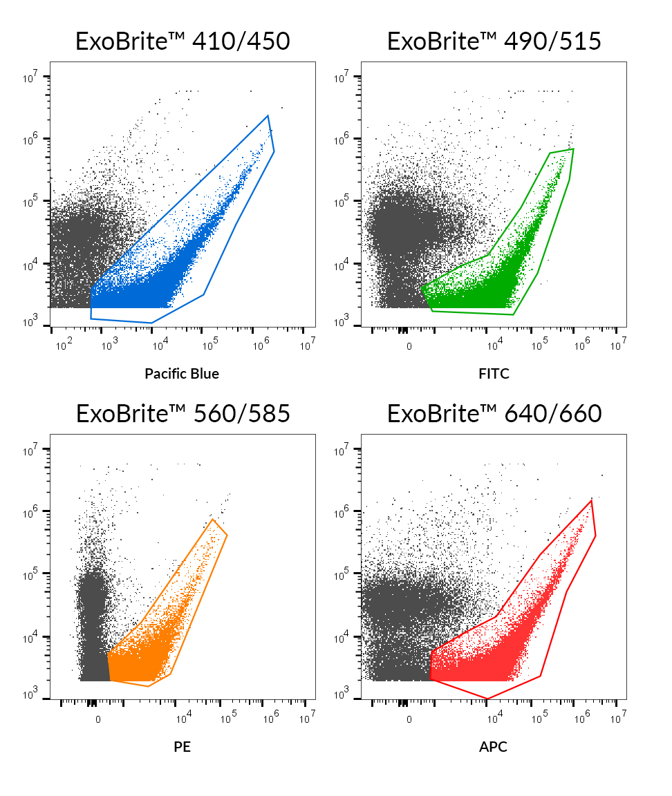
使用 ExoBrite™ EV 膜染色剂对 SEC 纯化的 MCF-7 衍生外泌体进行染色。
值得信赖的外泌体染色
ExoBrite™ EV 膜染色试剂盒设计用于通过流式细胞术或显微镜在最小背景下对分离的外泌体进行稳健染色。
特征
EV 和外泌体膜的明亮和特异性染色
适用于纯化的和珠粒结合的外泌体
针对流式细胞术检测进行了优化
兼容抗体共染
查看产品
ExoBrite™ EV Membrane Staining Kits
产品推荐
EvaGreen® Dye, 20X in Water
产品编号:31000-T
详细视图
TrueBlack® Lipofuscin Autofluorescence Quencher
产品编号:23007
详细视图

PMAxx™ Dye, 20 mM in H2O
产品编号:40069
详细视图
PMA (Propidium Monoazide)
产品编号:40019
详细视图
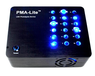
PMA-Lite™ LED Photolysis Device
产品编号:E90002
详细视图
PAGE GelRed® Nucleic Acid Gel Stain
产品编号:41008-T
详细视图